Bassett Collection of Stereoscopic Images of Human Anatomy
Exploration of liver, gall bladder, pancreas, duodenum and spleen
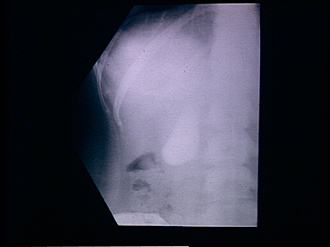
Gall bladder, normal cholangiogram
Image #147-1
KEYWORDS: Gallbladder, Liver, Pancreas, Spleen.
Creative Commons
Stanford holds the copyright to the David L. Bassett anatomical images and has assigned Creative Commons license Attribution-Share Alike 4.0 International to all of the images.
For additional information regarding use and permissions, please contact the Medical History Center.
Exploration of liver, gall bladder, pancreas, duodenum and spleen
Gall bladder, normal cholangiogram
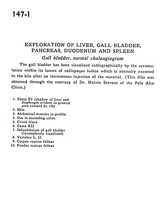
The gall bladder has been visualized radiographically by the accumulation within its lumen of radiopaque iodine which is normally excreted in the bile after the intravenous injection of the material. (This film was obtained through the courtesy of Dr. Melvin Stevens of the Palo Alto Clinic.)
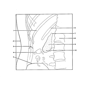
- Rib XI (shadow of liver and diaphragm evident in general area crossed by rib)
- Skin
- Abdominal muscles in profile
- Gas in ascending colon
- Iliac crest
- Rib XII
- Infundibulum of gall bladder (incompletely visualized)
- Vertebra L. II
- Body of gallbladder
- Fundus gallbladder


