Bassett Collection of Stereoscopic Images of Human Anatomy
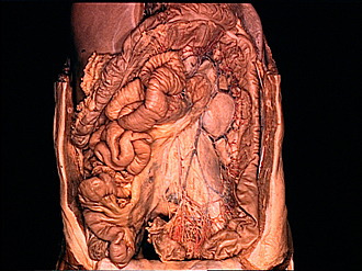
Dissection of jejunum, ileum and colon
Inferior mesenteric vessels and nerves, general view
Image #144-1
KEYWORDS: Large intestine, Peripheral nervous system, Small intestine, Vasculature, Overview.
Creative Commons
Stanford holds the copyright to the David L. Bassett anatomical images and has assigned Creative Commons license Attribution-Share Alike 4.0 International to all of the images.

For additional information regarding use and permissions, please contact the Medical History Center.
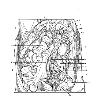
Dissection of jejunum, ileum and colon
Inferior mesenteric vessels and nerves, general view
The jejunum and ileum have been pulled to the right. The sigmoid mesocolon has been dissected. Lymphatic structures in this area are illustrated in view 141-4 ff. and 144-4.
- Transverse colon (elevated)
- Liver
- Jejunum (retracted)
- Root of mesenteries
- Ileum (retracted)
- Left colic flexure
- Spleen
- Left branch of middle colic artery
- Duodenojejunal flexure
- Kidney (covered by renal fascia)
- Descending colon
- Upper pointer: Left colic artery Lower pointer: Inferior mesenteric artery
- Psoas major muscle (covered by psoas fascia)
- Inferior mesenteric vein (pointers on tributary branches)
- Inferior mesenteric plexus
- Sigmoid artery
- Sigmoid colon
- Mesosigmoid colon (dissected)