Bassett Collection of Stereoscopic Images of Human Anatomy
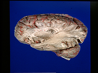
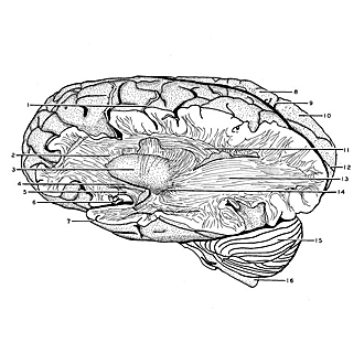
Exploration of the brain from its lateral aspect
External sagittal stratum
Image #14-2
KEYWORDS: Brain, Frontal lobe, Occipital lobe, Parietal lobe, Telencephalon, Temporal lobe.
Creative Commons
Stanford holds the copyright to the David L. Bassett anatomical images and has assigned Creative Commons license Attribution-Share Alike 4.0 International to all of the images.

For additional information regarding use and permissions, please contact the Medical History Center.
Exploration of the brain from its lateral aspect
External sagittal stratum
The dissection has been continued posteriorly to the occipital pole by the removal of cortex and association bundles including the posterior part of the superior longitudinal fasciculus. This exposes a massive system of fibers (the external sagittal stratum) running toward the occipital pole in the depths of the hemisphere. Many of these fibers interconnect thalamic and cortical centers and course in the retrolenticular and sublenticular parts of the internal capsule (e.g., posterior stalk of the thalamus, geniculocalcarine tract). Fibers of the visual radiation (geniculocalcarine tract) at first pass outward from the lateral geniculate body, in the sublenticular part of the internal capsule (14), then turn posteriorly. They apparently do not form any exaggerated forward loops as is sometimes depicted.
- Central sulcus (of Rolando)
- Frontal part internal capsule
- Lentiform nucleus
- Inferior occipitofrontal fasciculus (cut across)
- Uncinate fasciculus (cut across)
- Middle cerebral artery (cut off)
- Temporal pole
- Right parietal lobe
- Parieto-occipital fissure right
- Cuneus
- Superior longitudinal fasciculus (cut across)
- Calcarine fissure right
- External sagittal stratum
- Sublenticular part of internal capsule
- Cerebellum
- Spinal medulla