
Bassett Collection of Stereoscopic Images of Human Anatomy
Exploration of peritoneal cavity
Duodenojejunal junction; superior and inferior duodenal recesses
Image #139-3
KEYWORDS:
Creative Commons
Stanford holds the copyright to the David L. Bassett anatomical images and has assigned Creative Commons license Attribution-Share Alike 4.0 International to all of the images.

For additional information regarding use and permissions, please contact the Medical History Center.

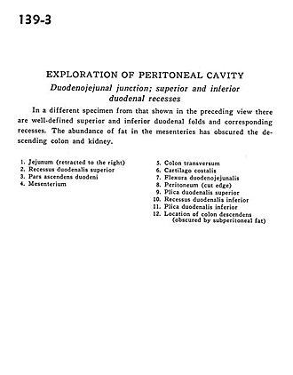
Exploration of peritoneal cavity
Duodenojejunal junction; superior and inferior duodenal recesses
In a different specimen from that shown in the preceding view there are well-defined superior and inferior duodenal folds and corresponding recesses. The abundance of fat in the mesenteries has obscured the descending colon and kidney.
- Jejunum (retracted to the right)
- Superior duodenal recess
- Ascending part of duodenum
- Mesenteries
- Transverse colon
- Costal cartilage
- Duodenojejunal flexure
- Peritoneum (cut edge)
- Superior duodenal fold
- Inferior duodenal recess
- Inferior duodenal fold
- Location of descending colon (obscured by subperitoneal fat)