Bassett Collection of Stereoscopic Images of Human Anatomy
Exploration of peritoneal cavity
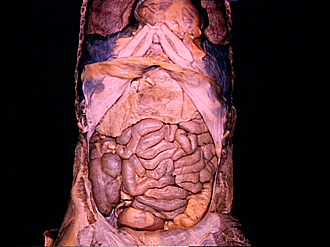
Abdominal organs in situ, general anterior view
Image #138-3
KEYWORDS: Overview.
Creative Commons
Stanford holds the copyright to the David L. Bassett anatomical images and has assigned Creative Commons license Attribution-Share Alike 4.0 International to all of the images.

For additional information regarding use and permissions, please contact the Medical History Center.
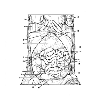

Exploration of peritoneal cavity
Abdominal organs in situ, general anterior view
The peritoneum has beem incised and reflected. The same specimen may be seen in a stage prior to the opening of the peritoneal sac by reference to view 135-2.
- Sternal xiphoid process
- Diaphragm
- Ligamentum teres (of liver) (exposed on inner aspect of reflected peritoneum note abundant extraperitoneal fat in adjacent area)
- Transverse colon
- Ascending colon
- Ileum
- Cecum
- Vermiform appendix
- Sigmoid colon
- Medial umbilical fold (exposed on inner aspect of reflected peritoneum)
- Median umbilical fold
- Heart
- Upper pointer: Stomach Lower pointer: Greater omentum
- Peritoneum (reflected)
- Jejunum
- External oblique
- Internal oblique
- Transversus abdominis muscle
- Descending colon