Bassett Collection of Stereoscopic Images of Human Anatomy
Dissection of female inguinal region
Femoral septum and associated lymph node, left side viewed from above
Image #138-1
KEYWORDS:
Creative Commons
Stanford holds the copyright to the David L. Bassett anatomical images and has assigned Creative Commons license Attribution-Share Alike 4.0 International to all of the images.

For additional information regarding use and permissions, please contact the Medical History Center.
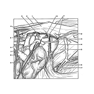
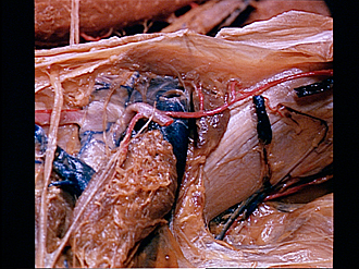
Dissection of female inguinal region
Femoral septum and associated lymph node, left side viewed from above
The specimen shown previously has been turned and is now viewed from above and inside. The pubic symphysis lies next to the right. Peritoneum and fascia have been removed to reveal the external iliac vessels (3,5) as they pass downward through the vascular compartment deep to the inguinal ligament (2). A thickened band of fascia known as the deep femoral arch, which is fused to the inguinal ligament has been elevated. The femoral septum (11) is visible medial to the external iliac vein. The cribriform nature of this septum is evident in the photograph. It forms the base of the femoral canal and is penetrated by lymphatic vessels which enter the lymphatic node (of Cloquet) (14) which is located just above the septum.
- Deep femoral arch (elevated)
- Inguinal ligament (elevated)
- External iliac vein
- Lymph vessel
- External iliac artery
- Deep circumflex iliac artery
- Inferior epigastric artery (cut off)
- Branch of femoral nerve
- External iliac lymph node
- Inferior epigastric vein (cut off)
- Femoral septum (pointer indicates approximate span of septum which closes base of femoral canal)
- Location of lacunar ligament (obscured by deep femoral arch)
- Pubic branch of inferior epigastric artery
- Iliac lymph node (of Cloquet)
- Superior ramus of pubic bone
- Endopelvic fascia (pointer on tab-like extension of this fascia into obturator foramen)
- Obturator artery (pointer on pubic branch)