
Bassett Collection of Stereoscopic Images of Human Anatomy
Dissection of female inguinal region
Inguinal canal (continued).
Image #137-5
KEYWORDS: Muscles and tendons, Peripheral nervous system.
Creative Commons
Stanford holds the copyright to the David L. Bassett anatomical images and has assigned Creative Commons license Attribution-Share Alike 4.0 International to all of the images.

For additional information regarding use and permissions, please contact the Medical History Center.
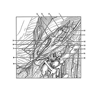

Dissection of female inguinal region
Inguinal canal (continued).
The transversus abdominis has been elevated along its lower border. The round ligament has been lifted away from the posterior wall of the inguinal canal.
- Transversus abdominis muscle
- Ligamentum teres (of uterus) (encased in fascial continuation from transversalis fascia)
- Internal oblique muscle (reflected)
- Inferior epigastric artery
- Conjoint tendon (falx inguinalis)
- Interfoveolar ligament (muscle fibers present)
- Branch of inferior epigastric artery
- Right pointer: Inguinal ligament Left pointer: Pubic tubercle
- Ligamentum teres (of uterus) (extending downward into labium majus)
- Internal oblique muscle (origin)
- Transversalis fascia
- Cremaster muscle (upper pointer indicates nerve supply, genital branch of genitofemoral nerve)
- Upper pointer: Inguinal ligament Lower pointer: Deep femoral arch (consisting of fibers associated with transversalis fascia)
- Superficial inguinal lymph node (retracted downward)
- Femoral sheath (pointer overlies femoral vein)
- Femoral canal (note lymphatic vessels passing through canal)