Bassett Collection of Stereoscopic Images of Human Anatomy
Dissection of anterolateral abdominal wall
General view of nerves and blood vessels of left rectus abdominis muscle
Image #134-5
KEYWORDS: Muscles and tendons, Peripheral nervous system, Vasculature, Overview.
Creative Commons
Stanford holds the copyright to the David L. Bassett anatomical images and has assigned Creative Commons license Attribution-Share Alike 4.0 International to all of the images.

For additional information regarding use and permissions, please contact the Medical History Center.


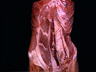
Dissection of anterolateral abdominal wall
General view of nerves and blood vessels of left rectus abdominis muscle
Both rectus muscles have been exposed within their sheaths. The left rectus has been reflected medially to expose its posterior surface
- External oblique muscle
- Right rectus abdominis muscle (exposed by removal of anterior layer of rectus sheath)
- Left rectus abdominis muscle (reflected medially)
- Inferior epigastric artery and vein
- Iliac crest
- Inguinal ligament
- Pyramidalis muscle
- Superior epigastric artery and vein
- Intercostal nerve VII
- Sheath of rectus abdominis muscle (posterior layer)
- Intercostal nerve X
- Transversus abdominis muscle
- Intercostal nerve XI
- Internal oblique muscle (muscle belly excised between areas indicated by pointers)
- Deep circumflex iliac artery (ascending branch)
- Transversalis fascia