
Bassett Collection of Stereoscopic Images of Human Anatomy
Dissection of anterolateral abdominal wall
General anterior view of external oblique, internal oblique and rectus abdominis muscles
Image #133-6
KEYWORDS: Muscles and tendons.
Creative Commons
Stanford holds the copyright to the David L. Bassett anatomical images and has assigned Creative Commons license Attribution-Share Alike 4.0 International to all of the images.

For additional information regarding use and permissions, please contact the Medical History Center.

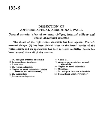
Dissection of anterolateral abdominal wall
General anterior view of external oblique, internal oblique and rectus abdominis muscles
The sheath of the right rectus abdominis has been opened. The left external oblique (9) has been divided close to the lateral border of the rectus sheath and its aponeurosis has been reflected medially. Fascia has been removed from all of the muscles.
- External oblique muscle
- Tendinous inscriptions
- Linea alba
- Rectus abdominis muscle
- Sheath of rectus abdominis muscle (anterior layer, cut and reflected)
- Pyramidalis muscle
- Inguinal ligament
- Rib VII
- Aponeurosis external oblique muscle
- Sheath of rectus abdominis muscle
- Umbilicus
- Internal oblique muscle
- Anterior superior iliac spine