
Bassett Collection of Stereoscopic Images of Human Anatomy
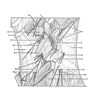
Dissection of anterolateral abdominal wall
Nerve supply to external oblique muscle (continued); fascia of internal oblique muscle
Image #133-5
KEYWORDS: Fascia, Muscles and tendons, Peripheral nervous system.
Creative Commons
Stanford holds the copyright to the David L. Bassett anatomical images and has assigned Creative Commons license Attribution-Share Alike 4.0 International to all of the images.

For additional information regarding use and permissions, please contact the Medical History Center.

Dissection of anterolateral abdominal wall
Nerve supply to external oblique muscle (continued); fascia of internal oblique muscle
The external oblique has been partially removed below its costal origins. The lower posterior part of the muscle has been dissected to display muscular branches of the twelfth thoracic nerve (subcostal n.) which enter the muscle from its internal aspect. Fascia between the external and internal oblique muscles have been preserved.
- Rib XII
- Rib XI
- External oblique muscle (origin)
- Costal cartilage X
- Lateral cutaneous branch intercostal nerve XI
- Posterior branches intercostal artery XI
- Internal oblique muscle (covered by fascia)
- Lateral cutaneous branch thoracic nerve XII (subcostal nerve)
- Muscular branches thoracic nerve XII
- External oblique muscle (cut across and reflected)
- Latissimus dorsi muscle (cut across)
- Thoracolumbar fascia
- Dorsal branch thoracic nerve XII (lateral branch)
- Aponeurosis of latissimus dorsi (in this specimen aponeurosis overlaps posterior border of external oblique muscle so that there is no lumbar triangle)
- Iliac crest
- Superior cluneal nerves
- Gluteus medius muscle