
Bassett Collection of Stereoscopic Images of Human Anatomy
Dissection of thorax from a posterior approach
Thoracic viscera.
Image #132-4
KEYWORDS: Diaphragm, Lung, Mediastinum, Muscles and tendons, Pleura.
Creative Commons
Stanford holds the copyright to the David L. Bassett anatomical images and has assigned Creative Commons license Attribution-Share Alike 4.0 International to all of the images.

For additional information regarding use and permissions, please contact the Medical History Center.
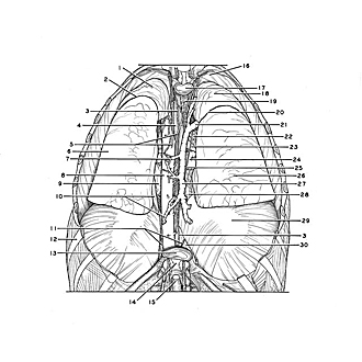

Dissection of thorax from a posterior approach
Thoracic viscera.
The posterior half of the costal pleura has been removed. The superior and posterior parts of the mediastinum have been dissected.
- Upper lobe left lung
- Oblique fissure
- Thoracic duct
- Esophagus
- Posterior intercostal artery II
- Lower lobe left lung
- Accessory hemiazygos vein
- Thoracic aorta (note origins of paired posterior intercostal arteries)
- Pericardium
- Upper pointer: Pulmonary ligament Lower pointer: Hemiazygos vein
- Greater splanchnic nerves (cut off)
- Rib IX
- Intervertebral disc Th. XI-XII
- Posterior longitudinal ligament
- Spinal cord
- Cupula pleurae
- Intervertebral disc Th. II- III
- Upper lobe right lung
- Trachea
- Oblique fissure
- Azygos vein (passing anteriorly to join superior vena cava)
- Right main bronchus
- Right pulmonary artery
- Right inferior pulmonary vein
- Right vagus nerve
- Inferior lobe right lung
- Azygos vein
- Pericardium enclosing inferior vena cava
- Diaphragm
- Mediastinal pleura