
Bassett Collection of Stereoscopic Images of Human Anatomy
Dissection of thorax from a posterior approach
Ligaments of costotransverse articulations in mid-thoracic region
Image #131-6
KEYWORDS: Bones joints cartilage, Fascia and connective tissue, Rib cage, Vertebral column.
Creative Commons
Stanford holds the copyright to the David L. Bassett anatomical images and has assigned Creative Commons license Attribution-Share Alike 4.0 International to all of the images.

For additional information regarding use and permissions, please contact the Medical History Center.
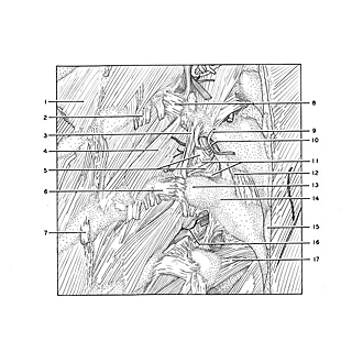
Dissection of thorax from a posterior approach
Ligaments of costotransverse articulations in mid-thoracic region
The dorsal muscles have been removed from the central area of the dissection.
- External intercostal muscle
- Longissimus thoracis muscle (tendon of insertion)
- Intertransverse ligament
- Levator costarum brevis muscle
- Superior costotransverse ligament (upper pointer on anterior division, lower pointer on posterior division)
- Costal tubercle VI
- Angle of rib VI (pointer on tendon of insertion of one slip of iliocostalis thoracis muscle)
- Lateral costotransverse ligament
- Dorsal branch posterior intercostal artery V
- Dorsal branch thoracic nerve V
- Upper pointer: Neck of rib Lower pointer: Costotransverse ligament (occupying foramen costotransversarium)
- Intervertebral joint capsule Th. V-VI
- Transverse process Th. vertebra VI
- Lamina of vertebral arch Th. VI
- Spinous process vertebrae Th. VI
- Nerve to levator costarum brevis muscle (branch of dorsal branch)
- Rotator brevis muscle