Bassett Collection of Stereoscopic Images of Human Anatomy
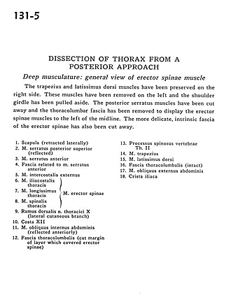
Dissection of thorax from a posterior approach
Deep musculature.
Image #131-5
KEYWORDS: Muscles and tendons.
Creative Commons
Stanford holds the copyright to the David L. Bassett anatomical images and has assigned Creative Commons license Attribution-Share Alike 4.0 International to all of the images.

For additional information regarding use and permissions, please contact the Medical History Center.
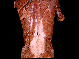
Dissection of thorax from a posterior approach
Deep musculature.
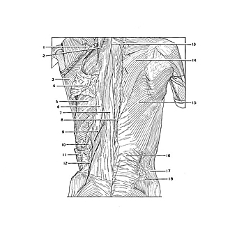
The trapezius and latissimus dorsi muscles have been preserved on the right side. These muscles have been removed on the left and the shoulder girdle has been pulled aside. The posterior serratus muscles have been cut away and the thoracolumbar fascia has been removed to display the erector spinae muscles to the left of the midline. The more delicate, intrinsic fascia of the erector spinae has also been cut away.
- Scapula (retracted laterally)
- Serratus posterior superior muscle (reflected)
- Serratus anterior muscle
- Fascia related to serratus anterior muscle
- External intercostal muscle
- Iliocostalis thoracis muscle
- Longissimus thoracis muscle
- Spinalis thoracis muscle (6-8 make up the erector spinae muscle)
- Dorsal branch thoracic nerve X (lateral cutaneous branch)
- Rib XII
- Internal abdominal oblique muscle (reflected anteriorly)
- Thoracolumbar fascia (cut margin of layer which covered erector spinae)
- Spinous process vertebrae Th. II
- Trapezius muscle
- Latissimus dorsi muscle
- Thoracolumbar fascia (intact)
- External abdominal oblique muscle
- Iliac crest