Bassett Collection of Stereoscopic Images of Human Anatomy
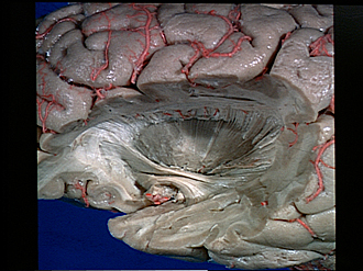
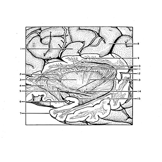
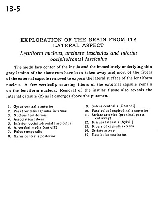
Exploration of the brain from its lateral aspect
Lentiform nucleus, uncinate fasciculus and inferior occipitofrontal fasciculus
Image #13-5
KEYWORDS: Brain, Telencephalon.
Creative Commons
Stanford holds the copyright to the David L. Bassett anatomical images and has assigned Creative Commons license Attribution-Share Alike 4.0 International to all of the images.

For additional information regarding use and permissions, please contact the Medical History Center.
Exploration of the brain from its lateral aspect
Lentiform nucleus, uncinate fasciculus and inferior occipitofrontal fasciculus
The medullary center of the insula and the immediately underlying thin gray lamina of the claustrum have been taken away and most of the fibers of the external capsule removed to expose the lateral surface of the lentiform nucleus. A few vertically coursing fibers of the external capsule remain on the lentiform nucleus. Removal of the insular tissue also reveals the internal capsule (2) as it emerges above the putamen.
- Precentral gyrus
- Frontal part internal capsule
- Lentiform nucleus
- Association fibers
- Inferior occipitofrontal fasciculus
- Middle cerebral artery (cut off)
- Temporal pole
- Postcentral gyrus
- Central sulcus (Rolandic)
- Superior longitudinal fasciculus
- Striate arteries (proximal parts cut away)
- Lateral fissure (Sylvian)
- Fibers of external capsule
- Striate artery
- Uncinate fasciculus