Bassett Collection of Stereoscopic Images of Human Anatomy
Dissection of mediastinum and paravertebral structures
Radiograph of esophagus, right anterior oblique view
Image #129-6
KEYWORDS: Bones joints cartilage, Esophagus, Vasculature, Vertebral column.
Creative Commons
Stanford holds the copyright to the David L. Bassett anatomical images and has assigned Creative Commons license Attribution-Share Alike 4.0 International to all of the images.

For additional information regarding use and permissions, please contact the Medical History Center.

Dissection of mediastinum and paravertebral structures
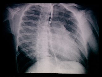
Radiograph of esophagus, right anterior oblique view
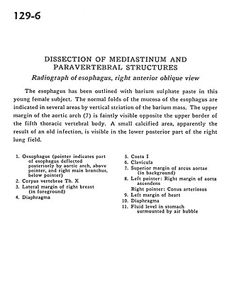
The esophagus has been outlined with barium sulfate paste in this young female subject. The normal folds of the mucosa of the esophagus are indicated in several areas by vertical striation of the barium mass. The upper margin of the aortic arch (7) is faintly visible opposite the upper border of the fifth thoracic vertebral body. A small calcified area, apparently the result of an old infection, is visible in the lower posterior part of the right lung field.
- Esophagus (pointer indicates part of esophagus deflected posteriorly by aortic arch, above pointer, and right main bronchus, below pointer)
- Body of vertebra Th. X
- Lateral margin of right breast (in foreground)
- Diaphragm
- Rib I
- Clavicle
- Superior margin of aortic arch (in background)
- Left pointer: Right margin of ascending aorta Right pointer: Conus arteriosus
- Left margin of heart
- Diaphragm
- Fluid level in stomach surmounted by air bubble