
Bassett Collection of Stereoscopic Images of Human Anatomy
Dissection of mediastinum and paravertebral structures
Esophageal plexus; pulmonary plexus; left recurrent laryngeal nerve
Image #129-3
KEYWORDS: Esophagus, Lung, Peripheral nervous system, Vasculature.
Creative Commons
Stanford holds the copyright to the David L. Bassett anatomical images and has assigned Creative Commons license Attribution-Share Alike 4.0 International to all of the images.

For additional information regarding use and permissions, please contact the Medical History Center.
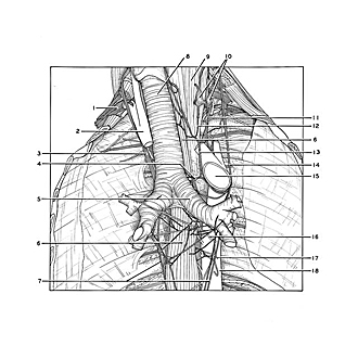

Dissection of mediastinum and paravertebral structures
Esophageal plexus; pulmonary plexus; left recurrent laryngeal nerve
The trachea and bronchi have been elevated. The aortic arch has been cut across and turned somewhat to the left to expose branches of the left recurrent laryngeal nerve.
- Brachial plexus
- Brachiocephalic trunk
- Vagus nerve right
- Tracheal and esophageal branches of recurrent laryngeal nerve
- Main bronchi
- Esophagus
- Esophageal plexus
- Trachea
- Longus colli muscle
- Stellate ganglion
- Sympathetic trunk
- Communication between stellate ganglion and recurrent laryngeal nerve
- Left recurrent laryngeal nerve
- Left vagus nerve
- Aortic arch (cut across)
- Bronchial branch of aorta
- Esophageal branch of aorta
- Thoracic aorta