
Bassett Collection of Stereoscopic Images of Human Anatomy
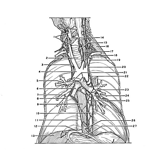
Dissection of mediastinum and paravertebral structures
Relations of tracheobronchial tree, aorta and esophagus, anterior view
Image #129-1
KEYWORDS: Esophagus, Lung, Mediastinum, Vasculature.
Creative Commons
Stanford holds the copyright to the David L. Bassett anatomical images and has assigned Creative Commons license Attribution-Share Alike 4.0 International to all of the images.

For additional information regarding use and permissions, please contact the Medical History Center.

Dissection of mediastinum and paravertebral structures
Relations of tracheobronchial tree, aorta and esophagus, anterior view
- Larynx
- Trachea
- Vagus nerve right
- Aortic arch
- Bronchus of upper right lobe
- Inferior tracheobronchial lymph nodes
- Bronchus of middle right lobe
- Right inferior lobe bronchus
- Esophagus
- Posterior mediastinal lymph node
- Vertebral column (covered by pleura)
- Inferior vena cava
- Diaphragm
- Vocal fold
- Esophagus (cut across near pharyngeal junction)
- Posterior scalene muscle
- Stellate ganglion
- Thoracic duct (cut off)
- Left subclavian artery
- Vagus nerve left
- Main bronchi
- Recurrent laryngeal nerve
- Left upper lobe bronchus
- Left lower lobe bronchus
- Thoracic aorta
- Phrenicoesophageal membrane
- Pericardium (remnant)