
Bassett Collection of Stereoscopic Images of Human Anatomy
Dissection of mediastinum and paravertebral structures
Upper thoracic part of right sympathetic trunk; intercostal vessels; azygos vein; right vagus nerve; pulmonary and esophageal plexuses
Image #128-4
KEYWORDS: Central nervous system, Peripheral nervous system, Vasculature.
Creative Commons
Stanford holds the copyright to the David L. Bassett anatomical images and has assigned Creative Commons license Attribution-Share Alike 4.0 International to all of the images.

For additional information regarding use and permissions, please contact the Medical History Center.
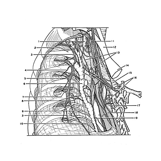
Dissection of mediastinum and paravertebral structures
Upper thoracic part of right sympathetic trunk; intercostal vessels; azygos vein; right vagus nerve; pulmonary and esophageal plexuses
The tracheobronchial tree has been pulled forward and to the left and the esophagus has been rotated slightly to the left.
- Posterior mediastinal lymph node
- Rib III
- Sympathetic trunk
- Ramus communicans (aberrant rami similar to this, which cross a rib to the next lower intercostal nerve, are present in several places on both sides of this specimen)
- Thoracic ganglion
- Azygos vein (cut off)
- Ramus communicans
- Lymph vessel
- Intercostal artery and vein VI
- Greater splanchnic nerve
- Vagus nerve right (several esophageal and tracheal branches visible along course of nerve)
- Brachiocephalic trunk
- Thoracic cardiac nerve (these nerves also send branches into pulmonary plexus)
- Aortic arch
- Trachea
- Pulmonary plexus
- Inferior tracheobronchial lymph node
- Esophagus
- Thoracic aorta