Bassett Collection of Stereoscopic Images of Human Anatomy
Dissection of mediastinum and paravertebral structures
Superior mediastinum.
Image #127-2
KEYWORDS: Mediastinum, Vasculature.
Creative Commons
Stanford holds the copyright to the David L. Bassett anatomical images and has assigned Creative Commons license Attribution-Share Alike 4.0 International to all of the images.

For additional information regarding use and permissions, please contact the Medical History Center.
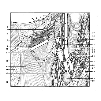
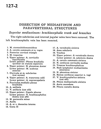
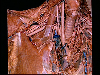
Dissection of mediastinum and paravertebral structures
Superior mediastinum.
The right subclavian and internal jugular veins have been removed. The left brachiocephalic vein has been resected.
- Sternocleidomastoid muscle
- Common carotid artery and vagus nerve
- Posterior cervical triangle
- Trapezius muscle
- Upper pointer: Superficial cervical artery Lower pointer: Brachial plexus (pointer on superior trunk)
- Upper pointer: Right phrenic nerve Lower pointer: Anterior scalene muscle
- Clavicle and subclavius muscle (cut across)
- Upper pointer: Transverse colli artery Lower pointer: Suprascapular artery
- Medial cord brachial plexus
- Axillary artery
- Axillary vein (cut off)
- Upper pointer: Cupula pleurae Lower pointer: Right brachiocephalic vein (cut oft)
- Pectoralis minor muscle
- Axilla
- Internal thoracic artery and vein
- Rib II
- Left vertebral artery
- Ansa subclavia
- Trachea
- Upper pointer: Right vertebral artery Lower pointer: Right subclavian artery
- Left common carotid artery
- Middle cervical cardiac nerve
- Brachiocephalic trunk
- Anterior mediastinal lymph nodes
- Left phrenic nerve
- Superior cardiac branch vagus nerve
- Left brachiocephalic vein (cut off)
- Left mediastinal pleura
- Ascending aorta
- Right costal pleura