
Bassett Collection of Stereoscopic Images of Human Anatomy
Dissection of mediastinum and paravertebral structures
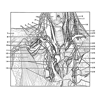
Superior mediastinum.
Image #127-1
KEYWORDS: Mediastinum, Vasculature.
Creative Commons
Stanford holds the copyright to the David L. Bassett anatomical images and has assigned Creative Commons license Attribution-Share Alike 4.0 International to all of the images.

For additional information regarding use and permissions, please contact the Medical History Center.

Dissection of mediastinum and paravertebral structures
Superior mediastinum.
The superior mediastinum has been opened by removing the manubrium and the upper part of the body of the sternum, together with parts of the first and second costal cartilages.
- Upper pointer: Thyroid cartilage (left lamina resected) Lower pointer: Cricoid cartilage
- Common carotid artery
- Internal jugular vein
- Anterior scalene muscle
- External jugular vein (cut across)
- Trapezius muscle and clavicle (cut off)
- Deltoid muscle
- Lymphatic duct right
- Right subclavian artery
- Upper pointer: Axillary artery Lower pointer: Axillary vein
- Subclavian trunk
- Upper pointer: Right bronchomediastinal trunk Lower pointer: Right brachiocephalic vein
- Pectoralis minor muscle
- Upper pointer: Superior vena cava Lower pointer: Costal pleura
- Sternal lymph nodes
- Vagus nerve left
- Vocal ligament
- Longus colli muscle
- Trachea
- Stellate ganglion
- Left subclavian artery
- Common carotid artery
- Left brachiocephalic vein
- Brachiocephalic trunk
- Thymus
- Costal pleura
- Internal thoracic artery and vein
- Ascending aorta (covered by fibrous layer of pericardium)
- Rib II