Bassett Collection of Stereoscopic Images of Human Anatomy
Dissection of lungs in situ
Left lung.
Image #125-6
KEYWORDS: Left lung, Lung, Peripheral nervous system, Vasculature.
Creative Commons
Stanford holds the copyright to the David L. Bassett anatomical images and has assigned Creative Commons license Attribution-Share Alike 4.0 International to all of the images.

For additional information regarding use and permissions, please contact the Medical History Center.

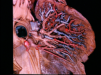
Dissection of lungs in situ
Left lung.
Branches of the left pulmonary artery have been cut away to reveal the more posterior subsegmental bronchi. The pulmonary plexus of nerves (5) has been more fully exposed. A branch of the bronchial artery (6) is visible.
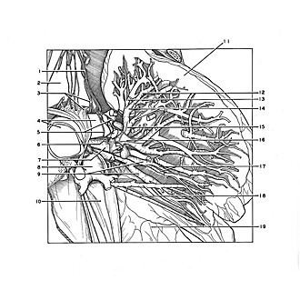
- Left vagus nerve
- Aortic arch
- Upper pointer: Recurrent laryngeal nerve Lower pointer: Ligamentum arteriosum
- Right pointer: Left pulmonary artery Left pointer: Pulmonary trunk
- Pulmonary plexus (note other unlabeled nerve filaments inferiorly)
- Bronchial branch of aorta
- Left upper lobe bronchus (note division into upper and lower parts)
- Left superior pulmonary vein (retracted medially)
- Upper pointer: Superior lingular bronchus Lower pointer: Inferior lingular bronchus
- Pericardium (retracted medially)
- Upper lobe left lung
- Anterior branch of apical subsegmental bronchus
- Apical branch of apical subsegmental bronchus
- Upper pointer: Apical subsegmental bronchus Lower pointer: Posterior subsegmental bronchus
- Posterior apical segmental bronchus
- Anterior segmental bronchus (note posterior branch close to origin of this bronchus)
- Superior lingular bronchus (upper pointer, anterior branch; lower pointer, posterior branch)
- Intersegmental lingular veins
- Lower lobe left lung