Bassett Collection of Stereoscopic Images of Human Anatomy
Dissection of lungs in situ
Left lung.
Image #125-5
KEYWORDS: Left lung, Lung, Lymphatics, Peripheral nervous system, Vasculature.
Creative Commons
Stanford holds the copyright to the David L. Bassett anatomical images and has assigned Creative Commons license Attribution-Share Alike 4.0 International to all of the images.

For additional information regarding use and permissions, please contact the Medical History Center.

Dissection of lungs in situ
Left lung.
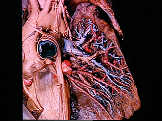
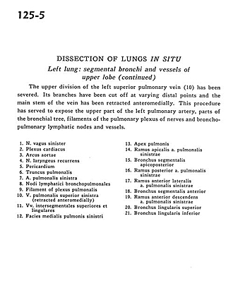
The upper division of the left superior pulmonary vein (10) has been severed. Its branches have been cut off at varying distal points and the main stem of the vein has been retracted anteromedially. This procedure has served to expose the upper part of the left pulmonary artery, parts of the bronchial tree, filaments of the pulmonary plexus of nerves and bronchopulmonary lymphatic nodes and vessels.
- Vagus nerve left
- Cardiac plexus
- Aortic arch
- Recurrent laryngeal nerve
- Pericardium
- Pulmonary trunk
- Left pulmonary artery
- Bronchopulmonary lymph nodes
- Filament of pulmonary plexus
- Left superior pulmonary vein (retracted anteromedially)
- Superior intersegmental and lingular veins
- Medial surface of left lung
- Apex of lung
- Apical branch left pulmonary artery
- Posterior apical segmental bronchus
- Branch posterior left pulmonary artery
- Anterior lateral branch left pulmonary artery
- Anterior segmental bronchus
- Anterior descending branch left pulmonary artery
- Superior lingular bronchus
- Inferior lingular bronchus