
Bassett Collection of Stereoscopic Images of Human Anatomy
Dissection of lungs in situ
Right lung.
Image #124-7
KEYWORDS: Left lung, Lung, Lymphatics.
Creative Commons
Stanford holds the copyright to the David L. Bassett anatomical images and has assigned Creative Commons license Attribution-Share Alike 4.0 International to all of the images.

For additional information regarding use and permissions, please contact the Medical History Center.

Dissection of lungs in situ
Right lung.
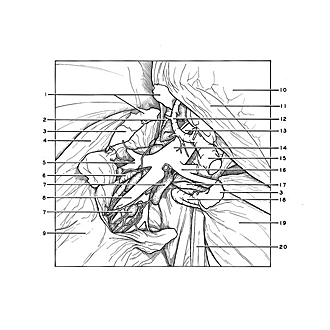
The right upper lobe (10) and middle lobe (19) have been pulled across the midline of the body to demonstrate the hilar region of the lower lobe and to display the interlobar parts of the pulmonary vessels. Small areas of fusion between the upper and lower lobes (1), and between the middle and lower lobes (3), have been divided in order to separate the lobes. The bronchopulmonary lymphatic vessels and nodes of the lower lobe have been preserved.
- Area of fusion between upper and middle lobes
- Right main bronchus
- Area of fusion between middle and lower lobes
- Interlobar surface lower lobe
- Superior branch of lower lobe, right pulmonary artery
- Superior segmental bronchus
- Bronchopulmonary lymph nodes
- Basilar part right pulmonary artery
- Lower lobe
- Upper lobe (reflected across midline)
- Interlobar surface upper lobe
- Right superior pulmonary vein (posterior branch to upper lobe)
- Right pulmonary artery (ascending branch of interlobar part)
- Right pulmonary artery (interlobar part)
- Right superior pulmonary vein (branch to middle lobe and anterior segment of upper lobe, cut off)
- Right pulmonary artery (branch to middle lobe)
- Bronchus of middle right lobe
- Right inferior pulmonary vein (branch to middle lobe)
- Middle lobe (pointer on interlobar surface)
- Pericardium