
Bassett Collection of Stereoscopic Images of Human Anatomy
Dissection of lungs in situ
Right lung.
Image #124-3
KEYWORDS: Lung, Right lung, Vasculature.
Creative Commons
Stanford holds the copyright to the David L. Bassett anatomical images and has assigned Creative Commons license Attribution-Share Alike 4.0 International to all of the images.

For additional information regarding use and permissions, please contact the Medical History Center.

Dissection of lungs in situ
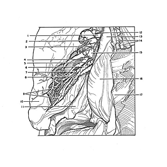
Right lung.
The close relation of branches of the pulmonary artery (4, blue) to the bronchial branches is evident, as well as the distinct intersegmental position of branches of the pulmonary vein (9, red).
- Right pulmonary artery (branches to superior lobe)
- Superior intersegmental vein
- Bronchopulmonary lymph node
- Branch to middle lobe pulmonary artery
- Filament of pulmonary plexus
- Medial segmental bronchus of middle lobe
- Superior branch of no. 6
- Inferior branch of no. 6
- Intersegmental veins of middle lobe
- Middle lobe right lung (mediastinal surface)
- Inferior lobe right lung
- Right vagus nerve
- Superior vena cava (covered by pleura)
- Internal thoracic artery and vein
- Right superior pulmonary vein
- Right atrium (covered by pleura and pericardium)
- Costal cartilage V