Bassett Collection of Stereoscopic Images of Human Anatomy
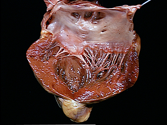
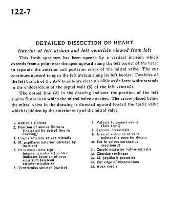
Detailed dissection of heart
Interior of left atrium and left ventricle viewed from left
Image #122-7
KEYWORDS: Heart, Left heart.
Creative Commons
Stanford holds the copyright to the David L. Bassett anatomical images and has assigned Creative Commons license Attribution-Share Alike 4.0 International to all of the images.

For additional information regarding use and permissions, please contact the Medical History Center.

Detailed dissection of heart
Interior of left atrium and left ventricle viewed from left
This fresh specimen has been opened by a vertical incision which extends from a point near the apex upward along the left border of the heart to separate the anterior and posterior cusps of the mitral valve. The cut continues upward to open the left atrium along its left border. Fascicles of the left branch of the A-V bundle are clearly visible as delicate white strands in the endocardium of the septal wall (5) of the left ventricle.
- Left auricle
- Position of anulus fibrosus (indicated by dotted line in drawing)
- Anterior cusp mitral valve
- Anterior papillary muscle (divided by incision)
- Muscular part interventricular septum (pointer indicates location of left crus atrioventricular bundle)
- Left ventricle (cavity)
- Valve of foramen ovale
- Interatrial septum
- Area of entrance of right superior pulmonary vein
- Fat in coronary sulcus (sectioned)
- Posterior cusp of mitral valve
- Chordae tendineae
- Posterior papillary muscle
- Cut edge of myocardium
- Apex of heart