Bassett Collection of Stereoscopic Images of Human Anatomy
Detailed dissection of heart
Interior of left ventricle.
Image #122-5
KEYWORDS: Heart, Left heart.
Creative Commons
Stanford holds the copyright to the David L. Bassett anatomical images and has assigned Creative Commons license Attribution-Share Alike 4.0 International to all of the images.

For additional information regarding use and permissions, please contact the Medical History Center.

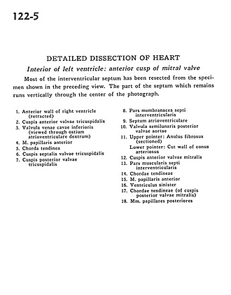
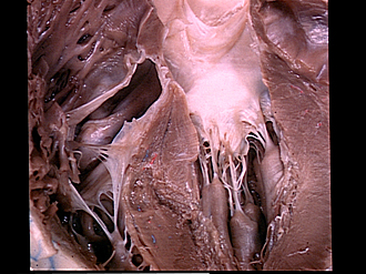
Detailed dissection of heart
Interior of left ventricle.
Most of the interventricular septum has been resected from the specimen shown in the preceding view. The part of the septum which remains runs vertically through the center of the photograph.
- Anterior wall of right ventricle (retracted)
- Anterior cusp tricuspid valve
- Valve of inferior vena cava (viewed through right atrioventricular opening)
- Anterior papillary muscle
- Chorda tendinea
- Septal (medial) cusp of tricuspid valve
- Posterior cusp tricuspid valve
- Membranous part interventricular septum
- Atrioventricular septum
- Posterior semilunar cusp (aortic valve)
- Upper pointer: Anulus fibrosus (sectioned) Lower pointer: Cut wall of conus arteriosus
- Anterior cusp of mitral valve
- Muscular part interventricular septum
- Chordae tendineae
- Anterior papillary muscle
- Left ventricle
- Chordae tendineae (of posterior cusp of mitral valve)
- Posterior papillary muscles