
Bassett Collection of Stereoscopic Images of Human Anatomy
Detailed dissection of heart
Atrial myocardium, basal view
Image #120-2
KEYWORDS: Heart, Left heart, Muscles and tendons, Right heart.
Creative Commons
Stanford holds the copyright to the David L. Bassett anatomical images and has assigned Creative Commons license Attribution-Share Alike 4.0 International to all of the images.

For additional information regarding use and permissions, please contact the Medical History Center.
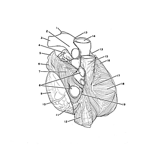
Detailed dissection of heart
Atrial myocardium, basal view
The epicardium has been removed from all parts of the atria with the exception of the left auricle.
- Ligamentum arteriosum
- Right pulmonary artery
- Left pulmonary artery
- Left auricle (epicardium intact)
- Left superior pulmonary vein
- Left inferior pulmonary vein
- Left atrium
- Upper pointer: Right superior pulmonary vein (note tributary joining vein close to atrial wall) Lower pointer: Right inferior pulmonary vein
- Left ventricle (epicardium intact)
- Posterior interventricular sulcus (pointer on middle cardiac vein)
- Right ventricle
- Inferior vena cava
- Pulmonary trunk
- Ascending aorta
- Superior vena cava
- Right auricle
- Sino-atrial node
- Right atrium
- Interatrial groove