
Bassett Collection of Stereoscopic Images of Human Anatomy
Exploration of the brain from its superior aspect
Lentiform nucleus, internal capsule, subthalamic nucleus
Image #12-5
KEYWORDS: Brain, Diencephalon, Telencephalon.
Creative Commons
Stanford holds the copyright to the David L. Bassett anatomical images and has assigned Creative Commons license Attribution-Share Alike 4.0 International to all of the images.

For additional information regarding use and permissions, please contact the Medical History Center.

Exploration of the brain from its superior aspect
Lentiform nucleus, internal capsule, subthalamic nucleus
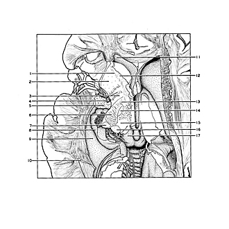
A horizontal cut has been made through the lentiform nucleus, internal capsule and thalamic area. The lamination within the lentiform nucleus is still demonstrable, although some of the nucleus was removed earlier. From the medial border of the globus pallidus fibers of the fasciculus lenticularis (13) pass through the internal capsule. The subthalamic nucleus (corpus Luysi) is cut through (14). The geniculate bodies are still present in this section which is cut lower than the previous view.
- Frontal part internal capsule
- Putamen
- Globus pallidus
- Anterior commissure (cut across)
- Amygdaloid nucleus (dissected)
- Internal capsule (cut horizontally)
- Lateral geniculate body
- Hippocampus
- Collateral eminence
- External sagittal stratum (cut across)
- Anterior horn lateral ventricle
- Head of caudate nucleus (dissected)
- Fasciculus lenticularis (fibers passing through internal capsule)
- Hypothalamic nucleus (corpus Luysi)
- Ventral posterior lateral nucleus of thalamus
- Medial geniculate body
- Superior colliculus