Bassett Collection of Stereoscopic Images of Human Anatomy
Exploration of the brain from its superior aspect
Inferior horn of lateral ventricle; hippocampus
Image #12-4
KEYWORDS: Brain, Telencephalon, Vasculature, Ventricules.
Creative Commons
Stanford holds the copyright to the David L. Bassett anatomical images and has assigned Creative Commons license Attribution-Share Alike 4.0 International to all of the images.

For additional information regarding use and permissions, please contact the Medical History Center.

Exploration of the brain from its superior aspect
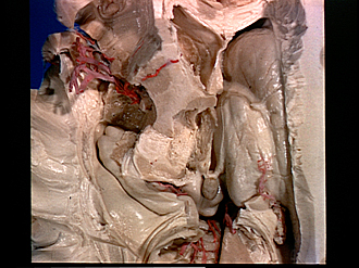
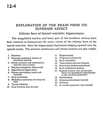
Inferior horn of lateral ventricle; hippocampus
The amygdaloid nucleus and lower part of the lentiform nucleus have been removed to demonstrate the entire extent of the inferior horn of the lateral ventricle. Note the hippocampal digitations bulging upward into the opened cavity. The anterior commissure and striate arteries are also visible.
- Putamen
- External medullary lamina of lentiform nucleus
- Striate arteries and anterior commissure (cut across)
- Amygdaloid nucleus (dissected)
- Hippocampal digitations
- Choroid plexus lateral ventricle
- Stria terminalis
- Medullary substance of temporal lobe
- Taenia fimbriae
- Fornix (crus) (cut across)
- Hippocampus
- Collateral trigone
- Stria terminalis
- Anterior tubercie of thalamus
- Mamillothalamic tract
- Stria medullaris thalami
- Internal capsule (cut across just above its downward continuation as the cerebral peduncle)
- Subthalamus
- Lateral geniculate body
- Pulvinar
- Posterior cerebral artery (cut across)