Bassett Collection of Stereoscopic Images of Human Anatomy
Detailed dissection of heart
Coronary vessels, posterior view
Image #119-6
KEYWORDS: Heart, Left heart, Right heart.
Creative Commons
Stanford holds the copyright to the David L. Bassett anatomical images and has assigned Creative Commons license Attribution-Share Alike 4.0 International to all of the images.

For additional information regarding use and permissions, please contact the Medical History Center.
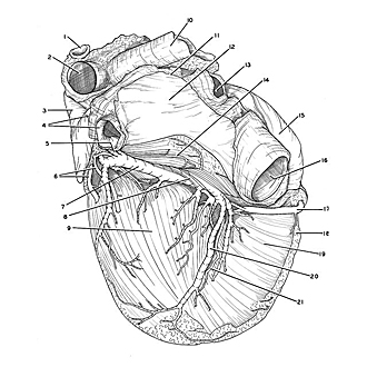

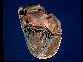
Detailed dissection of heart
Coronary vessels, posterior view
The specimen shown in the previous view has been dissected to demonstrate the distribution of the coronary arteries and cardiac veins on the posteroinferior aspect of the heart.
- Fragment of aorta at point of attachment of ligamentum arteriosum
- Left pulmonary artery
- Left auricle
- Left pulmonary veins (superior vein partially hidden)
- Left oblique atrial vein
- Ventricular branches of circumflex branch of left coronary artery
- Great cardiac vein
- Coronary sinus (covered by thin layer of muscle continuous with atrial myocardium)
- Left ventricle
- Right pulmonary artery
- Pericardial reflection at margin of oblique sinus
- Left atrium
- Right pulmonary veins
- Atrial branch of left coronary artery
- Right atrium
- Inferior vena cava
- Right coronary artery (in coronary sulcus)
- Margin of dissected. area
- Right ventricle
- Middle cardiac vein (in posterior interventricular sulcus)
- Posterior interventricular branch of right coronary artery