
Bassett Collection of Stereoscopic Images of Human Anatomy
Detailed dissection of heart
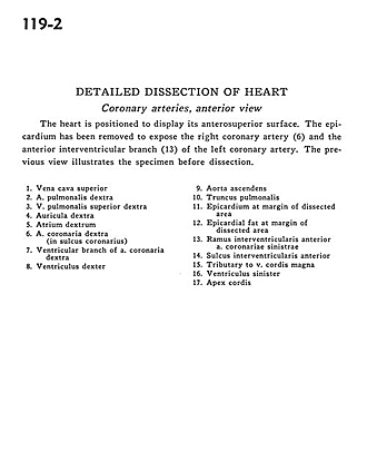
Coronary arteries, anterior view
Image #119-2
KEYWORDS: Heart, Left heart, Right heart, Vasculature.
Creative Commons
Stanford holds the copyright to the David L. Bassett anatomical images and has assigned Creative Commons license Attribution-Share Alike 4.0 International to all of the images.

For additional information regarding use and permissions, please contact the Medical History Center.

Detailed dissection of heart
Coronary arteries, anterior view
The heart is positioned to display its anterosuperior surface. The epicardium has been removed to expose the right coronary artery (6) and the anterior interventricular branch (13) of the left coronary artery. The previous view illustrates the specimen before dissection.
- Superior vena cava
- Right pulmonary artery
- Right superior pulmonary vein
- Right auricle
- Right atrium
- Right coronary artery (in coronary sulcus)
- Ventricular branch of right coronary artery
- Right ventricle
- Ascending aorta
- Pulmonary trunk
- Epicardium at margin of dissected area
- Epicardial fat at margin of dissected area
- Anterior interventricular branch of left coronary artery
- Anterior interventricular sulcus
- Tributary to great cardiac vein
- Left ventricle
- Apex of heart