
Bassett Collection of Stereoscopic Images of Human Anatomy
Dissection of pericardium and heart in situ
Interior of left atrium and left ventricle.
Image #118-5
KEYWORDS: Heart, Left heart, Pericardial sac.
Creative Commons
Stanford holds the copyright to the David L. Bassett anatomical images and has assigned Creative Commons license Attribution-Share Alike 4.0 International to all of the images.

For additional information regarding use and permissions, please contact the Medical History Center.
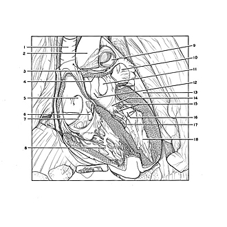
Dissection of pericardium and heart in situ
Interior of left atrium and left ventricle.
The latex cast has been cut away to display the cavity of the left atrium and the posterior cusp of the mitral valve with its associated chordae tendineae.
- Superior vena cava
- Ascending aorta
- Transverse pericardial sinus (indicated on drawing by double pointed arrow)
- Wall of left atrium (cut)
- Fossa ovalis
- Latex cast in coronary sinus
- Valve of inferior vena cava
- Right ventricle
- Pulmonary trunk
- Red latex cast in left auricle
- Latex cast protruding from left pulmonary veins
- Left atrium
- Pericardium
- Posterior cusp of mitral valve
- Chordae tendineae
- Anterior papillary muscle
- Posterior papillary muscle
- Left ventricle