
Bassett Collection of Stereoscopic Images of Human Anatomy
Dissection of pericardium and heart in situ
Interior of left ventricle.
Image #118-3
KEYWORDS: Heart, Left heart, Pericardial sac.
Creative Commons
Stanford holds the copyright to the David L. Bassett anatomical images and has assigned Creative Commons license Attribution-Share Alike 4.0 International to all of the images.

For additional information regarding use and permissions, please contact the Medical History Center.
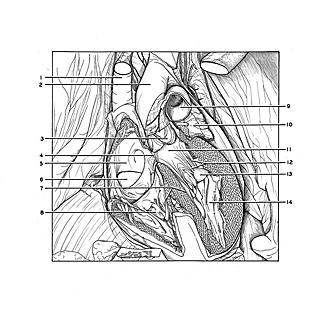
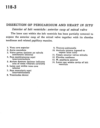
Dissection of pericardium and heart in situ
Interior of left ventricle.
The latex cast within the left ventricle has been partially removed to expose the anterior cusp of the mitral valve together with its chordae tendineae and related papillary muscles.
- Superior vena cava
- Ascending aorta
- Aortic valve (pointer on left semilunar cusp)
- Membranous part interventricular septum
- Right atrium (pointer indicates area of sinus venarum cavarum)
- Latex cast within inferior vena cava
- Muscular part of interventricular septum
- Right ventricle
- Pulmonary trunk
- Left auricle (opened to expose latex cast)
- Anterior cusp of mitral valve
- Chordae tendineae
- Anterior papillary muscle
- Latex cast within cavity of left ventricle