
Bassett Collection of Stereoscopic Images of Human Anatomy
Dissection of pericardium and heart in situ
Latex cast of cavity of left ventricle; septal branch of left coronary artery
Image #117-6
KEYWORDS: Heart, Left heart, Pericardial sac, Vasculature.
Creative Commons
Stanford holds the copyright to the David L. Bassett anatomical images and has assigned Creative Commons license Attribution-Share Alike 4.0 International to all of the images.

For additional information regarding use and permissions, please contact the Medical History Center.
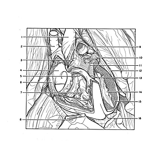
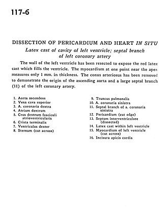
Dissection of pericardium and heart in situ
Latex cast of cavity of left ventricle; septal branch of left coronary artery
The wall of the left ventricle has been resected to expose the red latex cast which fills the ventricle. The myocardium at one point near the apex measures only 1 mm. in thickness. The conus arteriosus has been removed to demonstrate the origin of the ascending aorta and a large septal branch (11) of the left coronary artery.
- Ascending aorta
- Superior vena cava
- Right coronary artery
- Right atrium
- Right crus atrioventricular bundle
- Crista terminalis
- Right ventricle
- Sternum (cut across)
- Pulmonary trunk
- Left coronary artery
- Septal branch of left coronary artery
- Pericardium (cut edge)
- Interventricular septum (dissected)
- Latex cast within left ventricle
- Myocardium of left ventricle (cut across)
- Notch at apex of heart