Bassett Collection of Stereoscopic Images of Human Anatomy
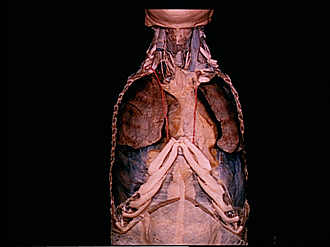
Thoracic viscera in situ
Anterior view of thoracic contents, rib cage removed
Image #116-3
KEYWORDS: Left lung, Lung, Pleura, Right lung, Overview.
Creative Commons
Stanford holds the copyright to the David L. Bassett anatomical images and has assigned Creative Commons license Attribution-Share Alike 4.0 International to all of the images.

For additional information regarding use and permissions, please contact the Medical History Center.


Thoracic viscera in situ
Anterior view of thoracic contents, rib cage removed
Both upper limbs have been detached. The upper nine ribs have been removed bilaterally with the exception of the first rib on the left. The manubrium and most of the body of the sternum have also been removed. The pleural cavities have been opened. On the right the apex of the lung is visible within the cupula of the pleura.
- Phrenic nerve
- Vagus nerve
- Right brachiocephalic vein (cut off)
- Fascia surrounding thymus
- Upper lobe right lung
- Pleura (cut along costomediastinal reflections)
- Middle lobe right lung
- Inferior lobe right lung
- Pleural fold
- Xiphoid process
- Sheath of rectus abdominis muscle (lamina posterior)
- Linea alba
- Rectus abdominis muscle
- Sheath of rectus abdominis muscle (lamina anterior)
- Thyroid cartilage
- Left internal jugular vein
- Thyroid gland
- Trachea
- Thymus
- Upper lobe left lung
- Internal thoracic artery
- Pericardium (covered by mediastinal pleura)
- Anterior mediastinal cavity
- Costal cartilage VI
- Diaphragm (covered by diaphragmatic pleura)
- Pleura (cut along costodiaphragmatic reflection)