Bassett Collection of Stereoscopic Images of Human Anatomy
Dissection of breast and anterolateral thoracic wall
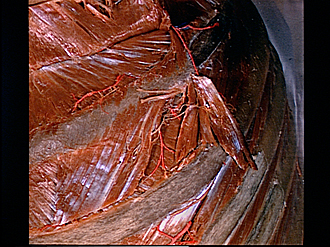
First and second left intercostal spaces, close-up view of nerves to external intercostal muscle
Image #115-4
KEYWORDS: Bones joints cartilage, Muscles and tendons, Peripheral nervous system, Rib cage.
Creative Commons
Stanford holds the copyright to the David L. Bassett anatomical images and has assigned Creative Commons license Attribution-Share Alike 4.0 International to all of the images.

For additional information regarding use and permissions, please contact the Medical History Center.


Dissection of breast and anterolateral thoracic wall
First and second left intercostal spaces, close-up view of nerves to external intercostal muscle
The external intercostal muscle has been reflected in the first intercostal space to expose the internal intercostal muscle. The intrinsic muscle fascia has been removed. In the second interspace the external intercostal has been dissected to illustrate a portion of its nerve supply.
- External intercostal muscle (reflected upward)
- Internal intercostal muscle
- Rib II
- External intercostal muscle
- Lateral cutaneous branch intercostal nerve II
- Nerve to external intercostal muscle
- Internal intercostal muscle
- Branch of posterior intercostal artery