
Bassett Collection of Stereoscopic Images of Human Anatomy
Sections of forearm and hand
Transverse section of right wrist
Image #112-3
KEYWORDS: Skin, Wrist.
Creative Commons
Stanford holds the copyright to the David L. Bassett anatomical images and has assigned Creative Commons license Attribution-Share Alike 4.0 International to all of the images.

For additional information regarding use and permissions, please contact the Medical History Center.

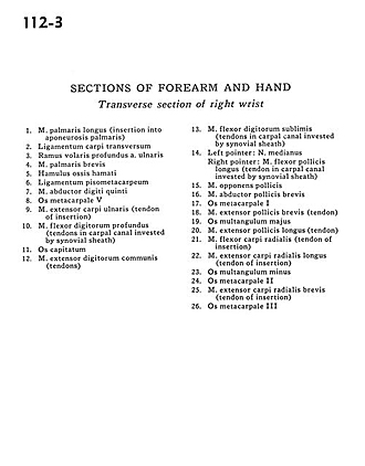
Sections of forearm and hand
Transverse section of right wrist
- Palmaris longus muscle (insertion into partial aponeurosis)
- Transverse carpal ligament
- Deep anterior branch ulnar artery
- Palmaris brevis muscle
- Hamulus of hamate bone
- Pisometacarpal ligament
- Abductor digiti minimi muscle
- Metacarpal V
- Extensor carpi ulnaris muscle (tendon of insertion)
- Flexor digitorum profundus muscle (tendons in carpal canal invested by synovial sheath)
- Capitate bone
- Common extensor digitorum muscle (tendons)
- Flexor digitorum superficialis (tendons in carpal canal invested by synovial sheath)
- Left pointer: Median nerve Right pointer: Flexor pollicis longus muscle (tendon in carpal canal invested by synovial sheath)
- Opponens pollicis muscle
- Abductor pollicis brevis muscle
- Metacarpal I
- Extensor pollicis brevis muscle (tendon)
- Trapezium bone
- Extensor pollicis longus muscle (tendon)
- Flexor carpi radialis muscle (tendon of insertion)
- Extensor carpi radialis longus muscle (tendon of insertion)
- Trapezoid bone
- Metacarpal II
- Extensor carpi radialis brevis muscle (tendon of insertion)
- Metacarpal III