Bassett Collection of Stereoscopic Images of Human Anatomy
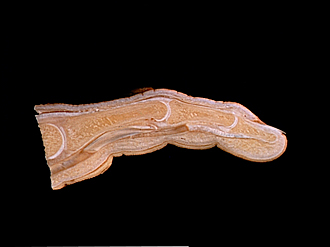
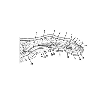
Sections of forearm and hand
Sagittal section of right middle finger, medial view
Image #111-6
KEYWORDS: Fascia ligaments and tendons, Hand and fingers, Muscles and tendons, Vasculature.
Creative Commons
Stanford holds the copyright to the David L. Bassett anatomical images and has assigned Creative Commons license Attribution-Share Alike 4.0 International to all of the images.

For additional information regarding use and permissions, please contact the Medical History Center.
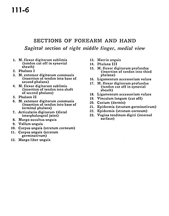
Sections of forearm and hand
Sagittal section of right middle finger, medial view
- Flexor digitorum superficialis (tendon cut off in synovial sheath)
- Phalanx I
- Common extensor digitorum muscle (insertion of tendon into base of second phalanx)
- Flexor digitorum superficialis (insertion of tendon into shaft of second phalanx)
- Phalanx II
- Common extensor digitorum muscle (insertion of tendon into base of terminal phalanx)
- Digital joint (distal interphalangeal joint)
- Hidden margin of nail
- Vallum of nail
- Body of nail (stratum corneum)
- Body of nail (stratum germinativum)
- Free margin of nail
- Matrix of nail
- Phalanx I
- Flexor digitorum profundus muscle (insertion of tendon into third phalanx)
- Anterior accessory ligament
- Flexor digitorum profundus muscle (tendon cut off in synovial sheath)
- Anterior accessory ligament
- Vinculum longum (cut off)
- Corium (dermis)
- Epidermis (stratum germinativum)
- Epidermis (stratum corneum)
- Tendon of digital sheath (internal surface)