
Bassett Collection of Stereoscopic Images of Human Anatomy
Joints of right index finger
Proximal interphalangeal joint opened, medial view
Image #111-5
KEYWORDS: Fascia ligaments and tendons, Hand and fingers, Muscles and tendons.
Creative Commons
Stanford holds the copyright to the David L. Bassett anatomical images and has assigned Creative Commons license Attribution-Share Alike 4.0 International to all of the images.

For additional information regarding use and permissions, please contact the Medical History Center.
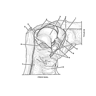

Joints of right index finger
Proximal interphalangeal joint opened, medial view
The ligaments and capsule of the joint have been cut. The extensor tendon and extensor expansion have also been transected to allow the proximal phalanx to be pulled away from the middle phalanx. The bones are in a position of flexion.
- Base of middle phalanx (covered by articular cartilage)
- Extensor expansion (divided)
- Phalangeal joint (covered by articular cartilage)
- Common extensor digitorum muscle (tendon of insertion divided)
- Joint capsule (cut)
- Collateral ligament (divided)
- Phalanx
- Synovial fold
- Anterior accessory ligament (divided)
- Ligament of digital sheath