Bassett Collection of Stereoscopic Images of Human Anatomy
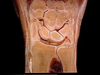
Joints of right wrist and hand
Frontal section, posterior view
Image #110-7
KEYWORDS: Fascia ligaments and tendons, Hand and fingers, Muscles and tendons, Wrist.
Creative Commons
Stanford holds the copyright to the David L. Bassett anatomical images and has assigned Creative Commons license Attribution-Share Alike 4.0 International to all of the images.

For additional information regarding use and permissions, please contact the Medical History Center.
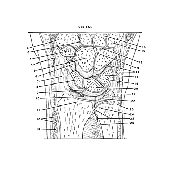
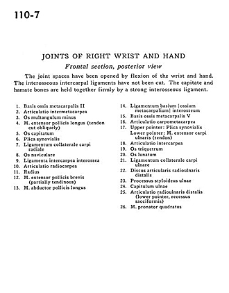
Joints of right wrist and hand
Frontal section, posterior view
The joint spaces have been opened by flexion of the wrist and hand. The interosseous intercarpal ligaments have not been cut. The capitate and hamate bones are held together firmly by a strong interosseous ligament.
- Base of metacarpal II
- Intermetacarpal joint
- Trapezoid bone
- Extensor pollicis longus muscle (tendon cut obliquely)
- Capitate bone
- Synovial fold
- Collateral carpi radial ligament
- Scaphoid bone
- Interosseous intercarpal ligament
- Radiocarpal joint
- Radius
- Extensor pollicis brevis muscle (partially tendinous)
- Abductor pollicis longus muscle
- Palmar metacarpal ligament
- Base of metacarpal V
- Carpometacarpal joint
- Upper pointer: Synovial fold Lower pointer: Extensor carpi ulnaris muscle (tendon)
- Intercarpal joint
- Triquetral bone
- Lunate bone
- Collateral carpi ulnar ligament
- Distal radioulnar articular disc
- Styloid process of ulna
- Head of ulna
- Distal radioulnar joint (lower pointer, sacciform recess)
- Pronator quadratus muscle