Bassett Collection of Stereoscopic Images of Human Anatomy
Lateral aspect of right hand
Nerve and blood supply to first dorsal interosseous muscle
Image #105-5
KEYWORDS: Hand and fingers, Neuralnetwork, Peripheral nervous system, Vasculature.
Creative Commons
Stanford holds the copyright to the David L. Bassett anatomical images and has assigned Creative Commons license Attribution-Share Alike 4.0 International to all of the images.

For additional information regarding use and permissions, please contact the Medical History Center.
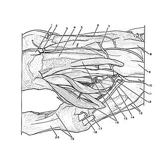
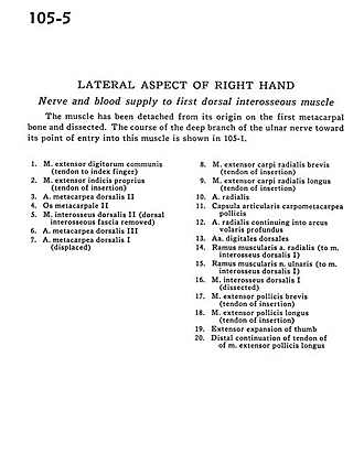
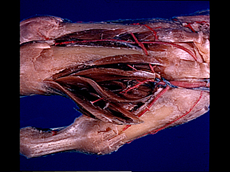
Lateral aspect of right hand
Nerve and blood supply to first dorsal interosseous muscle
The muscle has been detached from its origin on the first metacarpal bone and dissected. The course of the deep branch of the ulnar nerve toward its point of entry into this muscle is shown in 105-1.
- Common extensor digitorum muscle (tendon to index finger)
- Extensor indicis muscle (tendon of insertion)
- Dorsal metacarpal artery II
- Metacarpal II
- Dorsal interosseous muscle II (dorsal interosseous fascia removed)
- Dorsal metacarpal artery III
- Dorsal metacarpal artery I (displaced)
- Extensor carpi radialis brevis muscle (tendon of insertion)
- Extensor carpi radialis longus muscle (tendon of insertion)
- Radial artery
- Metacarpotrapezial joint capsule
- Radial artery continuing into deep palmar arch
- Dorsal digital arteries
- Muscular branch radial artery (to dorsal interosseous muscle I)
- Muscular branch of ulnar nerve (to dorsal interosseous muscle I)
- Dorsal interosseous muscle I (dissected)
- Extensor pollicis brevis muscle (tendon of insertion)
- Extensor pollicis longus muscle (tendon of insertion)
- Extensor expansion of thumb
- Distal continuation of tendon of of extensor pollicis longus muscle