
Bassett Collection of Stereoscopic Images of Human Anatomy
Volar aspect of right hand
Nerve supply to transverse head of adductor pollicis muscle
Image #104-6
KEYWORDS: Hand and fingers, Neuralnetwork, Vasculature.
Creative Commons
Stanford holds the copyright to the David L. Bassett anatomical images and has assigned Creative Commons license Attribution-Share Alike 4.0 International to all of the images.

For additional information regarding use and permissions, please contact the Medical History Center.
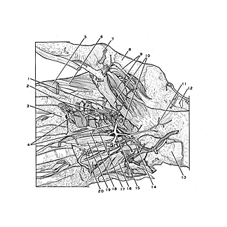
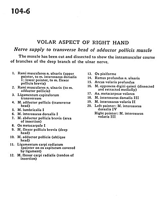
Volar aspect of right hand
Nerve supply to transverse head of adductor pollicis muscle
The muscle has been cut and dissected to show the intramuscular course of branches of the deep branch of the ulnar nerve.
- Muscular branches of ulnar nerve (upper pointer, to dorsal interosseous muscle I; lower pointer, to flexor pollicis brevis muscle)
- Muscular branches of ulnar nerve (to adductor pollicis muscle)
- Deep transverse metacarpal ligament
- Adductor pollicis muscle (transverse head)
- Lumbrical muscle I
- Dorsal interosseous muscle I
- Abductor pollicis brevis muscle (area of insertion)
- Metacarpal I
- Flexor pollicis brevis muscle (deep head)
- Adductor pollicis muscle (oblique head)
- Radiocarpal ligament (pointer on capitate bone covered by ligament)
- Flexor carpi radialis muscle (tendon of insertion)
- Pisiform bone
- Deep branch of ulnar nerve
- Deep palmar arch
- Opponens digiti minimi muscle (dissected and retracted medially)
- Anterior metacarpal arteries
- Dorsal interosseous muscle III
- Anterior interosseous muscle II
- Left pointer: Dorsal interosseous muscle IV Right pointer: Anterior interosseous muscle III