Bassett Collection of Stereoscopic Images of Human Anatomy
Volar aspect of right hand
Nerve supply to third and fourth lumbrical muscles and opponens digiti quinti muscle
Image #104-3
KEYWORDS: Fascia ligaments and tendons, Hand and fingers, Neuralnetwork, Vasculature.
Creative Commons
Stanford holds the copyright to the David L. Bassett anatomical images and has assigned Creative Commons license Attribution-Share Alike 4.0 International to all of the images.

For additional information regarding use and permissions, please contact the Medical History Center.


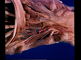
Volar aspect of right hand
Nerve supply to third and fourth lumbrical muscles and opponens digiti quinti muscle
The flexor tendons and lumbrical muscles have been retracted distally. The opponens digiti quinti muscle has been dissected.
- Sheath of common tendon of flexor muscles (opened, tendons and lumbrical muscles reflected distally)
- Lumbrical muscle III
- Lumbrical muscle IV
- Flexor digitorum profundus muscle (tendon to fifth finger)
- Lumbrical muscle I (cut off)
- Adductor pollicis muscle
- Flexor pollicis brevis muscle (deep head)
- Metacarpotrapezial joint capsule
- Transverse carpal ligament
- Carpal tunnel
- Branches of of ulnar nerve
- Ulnar artery
- Muscular branch ulnar artery (in position usually occupied by deep anterior branch ulnar artery)
- Deep branch of ulnar nerve
- Flexor and abductor digiti minimi muscles (area of origin)
- Muscular branch of ulnar nerve (to opponens digiti minimi muscle)
- Opponens digiti minimi muscle
- Deep anterior branch ulnar artery (aberrant origin more distal than usual)
- Anterior interosseous fascia
- Muscular branch of ulnar nerve (to Iumbrical muscle III)
- Proper palmar digital artery
- Muscular branch of ulnar nerve (to lumbrical muscle IV)
- Flexor and abductor digiti minimi muscles (area of insertion)
- Tendon of digital sheath V (opened)