
Bassett Collection of Stereoscopic Images of Human Anatomy
Volar aspect of right hand
Insertions of flexor tendons
Image #103-7
KEYWORDS: Fascia ligaments and tendons, Hand and fingers.
Creative Commons
Stanford holds the copyright to the David L. Bassett anatomical images and has assigned Creative Commons license Attribution-Share Alike 4.0 International to all of the images.

For additional information regarding use and permissions, please contact the Medical History Center.
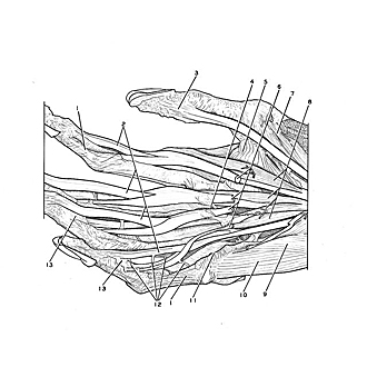
Volar aspect of right hand
Insertions of flexor tendons
The digital tendon sheaths have been opened and the flexor tendons elevated. The hand is viewed somewhat from its medial aspect.
- Heavy fascial bands extending along sides of fingers
- Flexor digitorum profundus muscle (tendon of insertion)
- Flexor pollicis longus muscle (insertion)
- Proximal limit of synovial sheath of third finger
- Adductor pollicis muscle (transverse head)
- Flexor digitorum superficialis (tendons of insertion)
- Flexor pollicis brevis muscle (deep head)
- Lumbrical muscles
- Flexor digiti minimi muscle
- Abductor digiti minimi muscle
- Ligament of digital sheath (cut edge)
- Vincula longa
- Vincula brevia