Bassett Collection of Stereoscopic Images of Human Anatomy
Volar aspect of right hand
Nerve supply to opponens pollicis muscle
Image #102-4
KEYWORDS: Hand and fingers, Neuralnetwork, Vasculature.
Creative Commons
Stanford holds the copyright to the David L. Bassett anatomical images and has assigned Creative Commons license Attribution-Share Alike 4.0 International to all of the images.

For additional information regarding use and permissions, please contact the Medical History Center.
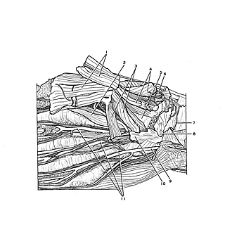
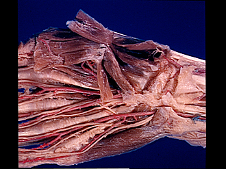
Volar aspect of right hand
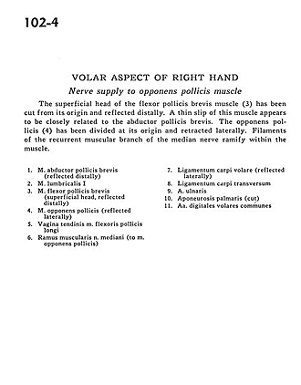
Nerve supply to opponens pollicis muscle
The superficial head of the flexor pollicis brevis muscle (3) has been cut from its origin and reflected distally. A thin slip of this muscle appears to be closely related to the abductor pollicis brevis. The opponens pollicis (4) has been divided at its origin and retracted laterally. Filaments of the recurrent muscular branch of the median nerve ramify within the muscle.
- Abductor pollicis brevis muscle (reflected distally)
- Lumbrical muscle
- Flexor pollicis brevis muscle (superficial head, reflected distally)
- Opponens pollicis muscle (reflected laterally)
- Tendon sheath of flexor pollicis longus muscle
- Muscular branch of median nerve (to opponens pollicis muscle)
- Volar carpal ligament (reflected laterally)
- Transverse carpal ligament
- Ulnar artery
- Palmar aponeurosis (cut)
- Common palmar digital arteries