Bassett Collection of Stereoscopic Images of Human Anatomy
Volar aspect of right hand
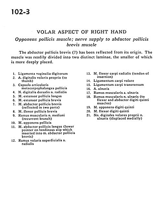
Opponens pollicis muscle; nerve supply to abductor pollicis brevis muscle
Image #102-3
KEYWORDS: Hand and fingers, Neuralnetwork, Vasculature.
Creative Commons
Stanford holds the copyright to the David L. Bassett anatomical images and has assigned Creative Commons license Attribution-Share Alike 4.0 International to all of the images.

For additional information regarding use and permissions, please contact the Medical History Center.
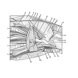
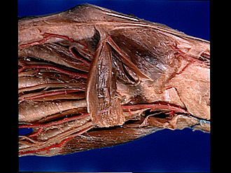
Volar aspect of right hand
Opponens pollicis muscle; nerve supply to abductor pollicis brevis muscle
The abductor pollicis brevis (7) has been reflected from its origin. The muscle was readily divided into two distinct laminae, the smaller of which is more deeply placed.
- Ligament of digital sheath
- Proper palmar digital artery (to thumb)
- Metacarpophalangeal pollicis joint capsule
- Dorsal digital nerve of radial nerve
- Extensor pollicis longus muscle
- Extensor pollicis brevis muscle
- Abductor pollicis brevis muscle (reflected in two parts)
- Flexor pollicis brevis muscle
- Muscular branch of median nerve (recurrent branch)
- Opponens pollicis muscle
- Abductor pollicis longus muscle (lower pointer on tendinous slip which inserted into abductor pollicis brevis muscle)
- Superficial anterior branch radial artery
- Flexor carpi radialis muscle (tendon of insertion)
- Anterior carpal ligament
- Transverse carpal ligament
- Ulnar artery
- Muscular branch ulnar artery
- Muscular branch of ulnar nerve (to flexor and abductor digiti minimi muscles)
- Opponens digiti minimi muscle
- Flexor digiti minimi muscle
- Proper palmar digital nerves of ulnar nerve (displaced medially)