Bassett Collection of Stereoscopic Images of Human Anatomy
Elbow joint
Articular cavity opened, posterior view
Image #100-6
KEYWORDS: Elbow, Muscles and tendons.
Creative Commons
Stanford holds the copyright to the David L. Bassett anatomical images and has assigned Creative Commons license Attribution-Share Alike 4.0 International to all of the images.

For additional information regarding use and permissions, please contact the Medical History Center.


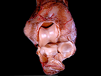
Elbow joint
Articular cavity opened, posterior view
The joint capsule and collateral ligaments have been cut. The radius and ulna have been pulled away from the humerus and flexed.
- Upper pointer: Cubital joint capsule (cut) Lower pointer: Synovial fold
- Medial epicondyle of humerus
- Trochlea of humerus
- Ulnar collateral ligament (divided)
- Synovial fold
- Olecranon
- Triceps brachii muscle (insertion)
- Olecranon fossa
- Lateral epicondyle of humerus
- Radial collateral ligament (divided)
- Head of humerus
- Fovea of head of radius
- Coronoid process of ulna
- Semilunar notch of ulna