
Bassett Collection of Stereoscopic Images of Human Anatomy
Elbow joint
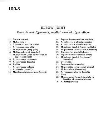
Capsule and ligaments, medial view of right elbow
Image #100-3
KEYWORDS: Elbow, Fascia ligaments and tendons, Muscles and tendons.
Creative Commons
Stanford holds the copyright to the David L. Bassett anatomical images and has assigned Creative Commons license Attribution-Share Alike 4.0 International to all of the images.

For additional information regarding use and permissions, please contact the Medical History Center.

Elbow joint
Capsule and ligaments, medial view of right elbow
- Body of humerus
- Brachialis muscle
- Cubital joint capsule
- Recurrent radial artery
- Supinator muscle (deep part)
- Biceps brachii muscle (tendon)
- Supinator muscle (area of insertion of superficial part)
- Recurrent interosseous artery
- Dorsal interosseous artery
- Radius
- AnteriOr interosseous artery
- Ulnar artery (cut off)
- Antebrachial interosseous membrane
- Medial intermuscular septum:
- Superior ulnar collateral artery
- Inferior ulnar collateral artery
- Triceps brachii muscle (medial head)
- Pronator teres muscle (humeral head)
- Medial epicondyle of humerus
- Ulnar collateral ligament
- Triceps brachii muscle (tendon of insertion)
- Olecranon
- Common flexor tendon
- Pronator teres muscle (ulnar head)
- Brachialis muscle (insertion)
- Dorsal recurrent ulnar artery
- Ulna
- Supinator muscle (muscle fascicle in location of chorda obliqua)
- Nutritive artery of ulna