Bassett Collection of Stereoscopic Images of Human Anatomy
Exploration of the brain from its superior aspect
Relations of the fornix, septum pellucida and lateral ventricles
Image #10-5
KEYWORDS: Brain, Diencephalon, Telencephalon, Ventricules.
Creative Commons
Stanford holds the copyright to the David L. Bassett anatomical images and has assigned Creative Commons license Attribution-Share Alike 4.0 International to all of the images.

For additional information regarding use and permissions, please contact the Medical History Center.


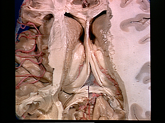
Exploration of the brain from its superior aspect
Relations of the fornix, septum pellucida and lateral ventricles
The central portion of the corpus callosum is now entirely removed to reveal the paired lateral ventricles separated by the septum pellucidum. The relations of the caudate nuclei, laminae affixae and crura and body of the fornix are discernible. The ependymal covering of the left caudate nucleus has been removed. On the right the ependyma is intact and just beneath it a number of uninjected veins are present. These are tributary to the vena terminalis.
- Corona radiata (dissected)
- Corpus callosum (near genu)
- Head of caudate nucleus
- Anterior tubercle of thalamus
- Superior longitudinal fasciculus (cut across)
- Occipital part corona radiata
- Tapetum (cut across)
- Cingulum (cut across)
- Anterior horn lateral ventricle
- Septum pellucidum (cut across at line of attachment to corpus callosum)
- Position of interventricular foramen (of Monro)
- Central part lateral ventricle
- Position of stria terminalis and vena terminalis (coursing parallel to caudate nucleus)
- Fornix (body)
- Remnant of choroid plexus lateral ventricle
- Lamina affixa
- Meninges within transverse fissure
- Hippocampus