Bassett Collection of Stereoscopic Images of Human Anatomy
Exploration of the meninges and brain in situ
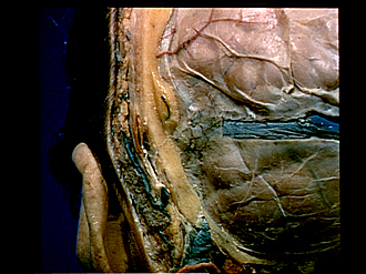
Close-up view of layers of scalp, calvaria and dura mater in left posterior auricular region
Image #1-6
KEYWORDS: Bones cartilage joints, Brain, Meninges, Scalp, Overview.
Creative Commons
Stanford holds the copyright to the David L. Bassett anatomical images and has assigned Creative Commons license Attribution-Share Alike 4.0 International to all of the images.

For additional information regarding use and permissions, please contact the Medical History Center.

Exploration of the meninges and brain in situ
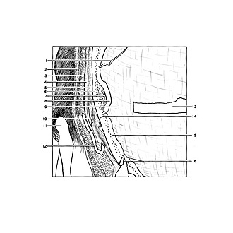
Close-up view of layers of scalp, calvaria and dura mater in left posterior auricular region
- Middle meningeal artery (posterior branch)
- Skin
- Superficial fascia
- Galea aponeurotica
- Pericranium
- External plate of occipital bone
- Posterior temporal diploic vein
- Internal plate of occipital bone
- Dura mater (note plexus of small dural veins in this area in the view)
- Occipitalis muscle (cut across)
- Auricle
- Posterior auricular vein
- Transverse sinus (dural covering cut away)
- Parietomastoid suture
- Mastoid part of temporal bone
- Mastoid emissary and occipitomastoid suture